大肠杆菌载体 E.coli Vector 大肠杆菌宿主菌株 E.coli 细菌广宿主载体 bacateria broad range host vector 链霉菌载体及菌株 Streptomyces 芽孢杆菌载体 Bacillus vector 芽孢杆菌宿主菌株 乳酸菌载体 lactic acid bacteria vector 乳酸菌宿主菌株 lactic acid bacteria strain 细菌基因敲除载体 毕赤酵母载体 毕赤酵母宿主菌株 酿酒酵母载体 酿酒酵母宿主菌株 丝状真菌载体 mold/fungi vector 乳酸克鲁维酵母载体 酵母真菌基因敲除基因编辑载体 植物细胞载体 plant cell vector 农杆菌菌株Agrobacterium tumefaciens strain 植物细胞基因敲除载体 plant cell 哺乳动物细胞载体 哺乳动物细胞荧光载体 荧光素酶报告基因载体 哺乳动物细胞基因敲除基因编辑载体 杂交系统 慢病毒载体 腺病毒载体 逆转录病毒载体 杆状病毒表达载体 基因干扰 RNAi载体 基因/cDNA/ORF 转座子质粒系统 transposon 金黄色葡萄球菌载体 staphylococcus aureus 假单胞菌载体 噬菌体 phage 不动杆菌载体 双岐杆菌载体 藻类表达载体 链球菌载体 厌氧菌载体 基因治疗载体 大肠杆菌基因突变体菌株 细菌荧光质粒 白色念珠菌载体 体外转录载体 谷氨酸棒杆菌载体 酿酒酵母基因突变体菌株 线虫载体 斑马鱼载体 Zebra fish 果蝇,昆虫载体Drosophila 鱼类细胞载体 fish cell 分支杆菌载体 克雷伯菌 枯草芽孢杆菌基因缺失突变株 基因ORF 金黄色葡萄球菌基因敲除突变株 肺炎克雷伯菌基因敲除突变株
| 近红外荧光蛋白-特别适合活体成像的一种荧光蛋白 |
| 发布时间:2021-11-06 22:42:44 | 浏览次数: |
Science:新近红外荧光蛋白让我们能观察内部器官
叶史瓦大学Albert Einstein医学院研究人员首次研制出独特的荧光蛋白,科学家借助该蛋白质可清晰地观察活体的内部器官,因而摆脱外科手术的解剖刀和有辐射风险的成像技术。
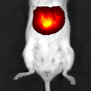
肝细胞表达iRFP蛋白 这一新探针在全身成像领域具有突破性意义,它能让医生在监视肿瘤生长过程中不留下创伤,因而对癌症治疗效果的评估有积极作用。与人体扫描技术相比,荧光蛋白成像技术不涉及辐射风险和对比剂。研究结果发表在7月17日 Nature Biotechnology期刊的网络版上。 过去的20年,科学家尝试了水母和珊瑚的各种有色荧光蛋白用于显示细胞、细胞器和分子,但是荧光探针在哺乳动物体内的使用却存在挑战,动物血液中血红蛋白能高效地吸收蓝光、绿光、红光以及其它波长的光。荧光蛋白的激发光和荧光蛋白“亮起来”后的发射光都会受到血红蛋白的吸收干扰。 Einstein医学院解剖与结构生物学副教授 Vladislav Verkhusha博士为了解决这一问题,他的实验室从细菌光敏色素(用于检测光)提取出荧光蛋白,这一荧光蛋白(称为iRFP)能吸收和发射电磁频场的近红外光,该频段的光对于哺乳动物组织而言几乎是透明的。 研究人员把荧光蛋白定位在肝脏中,这一器官由于血液含量高很难被观察。 含有iRFP基因的腺病毒颗粒注入小鼠体内。一旦载有iRFP基因的病毒感染肝细胞,感染细胞会表达该基因并合成iRFP蛋白质。接着,小鼠暴露在近红外光下,全身成像仪能观察到发射荧光的肝脏。最早在感染后的第2天检测出肝脏发荧光,5天后,荧光强度达到峰值。附加实验表明,iRFP蛋白质对哺乳动物没有毒害作用。 论文的第1作者Grigory Filonov博士是 Einstein 医学院 Verkhusha 实验室的一个成员。他说,据研究所知,iRFP蛋白在活体肝脏显示方面优于其它荧光蛋白,它不仅产生非常明亮的图像,而且长时期内保持很高的稳定性。我们相信它将在全身无创成像领域具有广阔的应用。 Filonov博士还说,与标准x-光和CT扫描相比,荧光蛋白成像没有辐射风险。此外,iRFP 蛋白成像很清晰,不需要对比剂,而核磁共振成像(MRI)需要吞下或注射对比剂以使得机体内部结构更清晰。(生物探索译 Pobee)
生物探索推荐英文原文 Newly Developed Fluorescent Protein Makes Internal Organs Visible Researchers at Albert Einstein College of Medicine of Yeshiva University have developed the first fluorescent protein that enables scientists to clearly "see" the internal organs of living animals without the need for a scalpel or imaging techniques that can have side effects or increase radiation exposure. The new probe could prove to be a breakthrough in whole-body imaging -- allowing doctors, for example, to noninvasively monitor the growth of tumors in order to assess the effectiveness of anti-cancer therapies. In contrast to other body-scanning techniques, fluorescent-protein imaging does not involve radiation exposure or require the use of contrast agents. The findings are described in the July 17 online edition of Nature Biotechnology. For the past 20 years, scientists have used a variety of colored fluorescent proteins, derived from jellyfish and corals, to visualize cells and their organelles and molecules. But using fluorescent probes to peer inside live mammals has posed a major challenge. The reason: hemoglobin in an animal's blood effectively absorbs the blue, green, red and other wavelengths used to stimulate standard fluorescent proteins along with any wavelengths emitted by the proteins when they do light up. To overcome that roadblock, the laboratory of Vladislav Verkhusha, Ph.D., associate professor of anatomy and structural biology at Einstein and the study's senior author, engineered a fluorescent protein from a bacterial phytochrome (the pigment that a species of bacteria uses to detect light). This new phytochrome-based fluorescent protein, dubbed iRFP, both absorbs and emits light in the near-infrared portion of the electromagnetic spectrum- the spectral region in which mammalian tissues are nearly transparent. The researchers targeted their fluorescent protein to the liver -- an organ particularly difficult to visualize because of its high blood content. Adenovirus particles containing the gene for iRFP were injected into mice. Once the viruses and their gene cargoes infected liver cells, the infected cells expressed the gene and produced iRFP protein. The mice were then exposed to near-infrared light and it was possible to visualize the resulting emitted fluorescent light using a whole-body imaging device. Fluorescence of the liver in the infected mice was first detected the second day after infection and reached a peak at day five. Additional experiments showed that the iRFP fluorescent protein was nontoxic. "Our study found that iRFP was far superior to the other fluorescent proteins that reportedly help in visualizing the livers of live animals," said Grigory Filonov, Ph.D., a postdoctoral fellow in Dr. Verkhusha''''s laboratory at Einstein, and the first author of the Nature Biotechnology paper. "iRFP not only produced a far brighter image, with higher contrast than the other fluorescent proteins, but was also very stable over time. We believe it will significantly broaden the potential uses for noninvasive whole-body imaging." Dr. Filonov noted that fluorescent-protein imaging involves no radiation risk, which can occur with standard x-rays and computed tomography (CT) scanning. And unlike magnetic resonance imaging (MRI), in which contrasting agents must sometimes be swallowed or injected to make internal body structures more visible, the contrast provided by iRFP is so vibrant that contrasting agents are not needed. |
| 上一篇:地衣芽孢杆菌感受态制备和电转化方法 下一篇:CRISPR-Cas9技术简介 |